
The Benefits of Routine Mammography: A Comprehensive Guide from Novocare

What to Expect from Novocare’s Comprehensive CT-Scan Services
September 12, 2025
How OPG X-Rays Are Used in Dentistry: Novocare’s Diagnostic Services
September 12, 2025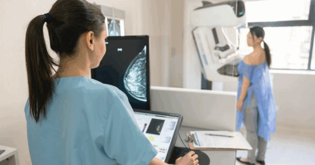
Introduction
Breast cancer is one of the most common cancers among women worldwide. Early detection is crucial to improving survival rates and ensuring effective treatment. Routine mammography is a key preventive tool that helps detect breast abnormalities even before symptoms appear.
At Novocare Hospital, we provide advanced mammography services with a focus on accuracy, safety, and patient comfort. This guide explores the benefits of routine mammography, how it works, who should get screened, and what to expect during the procedure.
1. What is Mammography?
Mammography is a specialized medical imaging technique that uses low-dose X-rays to examine the breast tissue. It is a vital part of women’s health check-ups and plays a central role in early breast cancer detection.
How Mammography Works
- The breast is placed on a flat surface of the mammography machine.
- Gentle compression spreads the tissue for clear imaging.
- Low-dose X-rays capture detailed images of internal breast structures.
- Radiologists analyze the images to detect lumps, cysts, or microcalcifications.
Types of Mammography at Novocare:
- Digital Mammography: High-resolution digital images for precise diagnosis.
- 3D Mammography (Tomosynthesis): Multi-layered images improve detection accuracy.
- Diagnostic Mammography: Focused scans for patients with symptoms or abnormal screening results.
2. Who Should Get Routine Mammography?
Routine mammography is recommended for women at various risk levels.
General Guidelines:
- Women aged 40–49: Discuss personalized screening based on risk factors.
- Women aged 50–74: Recommended every 1–2 years.
- High-risk individuals: Women with family history, genetic mutations (BRCA1/BRCA2), or previous breast conditions may require earlier and more frequent screening.
Risk Factors That Increase Screening Needs:
- Family history of breast cancer
- Genetic predisposition (BRCA mutations)
- Hormonal factors (early menstruation, late menopause)
- Previous breast biopsies or abnormal findings
3. Benefits of Routine Mammography
Routine mammography provides several critical benefits that significantly improve breast health outcomes.
Key Benefits:
- Early Detection of Breast Cancer: Identifies cancers before symptoms appear, improving survival rates.
- Reduced Mortality: Early-stage detection allows for less aggressive treatment and higher recovery chances.
- Detection of Non-Cancerous Conditions: Identifies cysts, calcifications, and benign tumors, enabling monitoring.
- Peace of Mind: Regular screenings reduce anxiety about breast health.
- Personalized Care: Helps healthcare providers tailor treatment plans based on findings.
Table: Mammography Benefits by Outcome
| Outcome | Benefit |
|---|---|
| Early Cancer Detection | Higher survival rates and better prognosis |
| Reduced Mortality | Timely treatment reduces advanced-stage breast cancer risks |
| Benign Condition Detection | Monitoring and preventive care |
| Informed Treatment Planning | Helps in planning surgery, chemotherapy, or radiation if needed |
| Patient Reassurance | Routine checks provide peace of mind and proactive health management |
4. What to Expect During a Mammography Appointment
Understanding the procedure can ease anxiety and make the process smoother.
Step-by-Step Process:
- Check-In: Confirm personal and medical information.
- Preparation: Remove clothing and jewelry from the waist up; wear a gown provided by the clinic.
- Positioning: A trained technician positions the breast on the mammography plate.
- Compression: Gentle compression spreads breast tissue for clear imaging.
- Image Capture: X-rays are taken from different angles.
- Completion: The procedure typically lasts 15–30 minutes.
- Result Review: Radiologists analyze the images and share findings with your physician.
Tips for Comfort:
- Schedule the mammogram one week after your period to reduce breast tenderness.
- Avoid deodorants or powders on the day of the scan, as they may affect image quality.
- Communicate any discomfort to the technician; compression is brief.
5. Advanced Technology at Novocare
Novocare’s state-of-the-art mammography equipment ensures accurate and comfortable imaging:
- Digital Mammography: Provides high-resolution images for detailed analysis.
- 3D Tomosynthesis: Captures layered images for better detection in dense breast tissue.
- Computer-Aided Detection (CAD): Assists radiologists in identifying abnormalities.
- Low Radiation Dose: Prioritizes patient safety without compromising image quality.
6. Common Myths About Mammography
There are misconceptions that may prevent women from getting screened.
- Myth 1: Mammograms are painful.
Fact: Slight discomfort may occur, but it is brief, and technicians minimize discomfort. - Myth 2: Only women with symptoms need a mammogram.
Fact: Routine screening is essential even if no symptoms are present. - Myth 3: Mammograms cause cancer.
Fact: Radiation exposure is very low, and the benefits far outweigh the minimal risk. - Myth 4: Breast cancer is always hereditary.
Fact: Most breast cancers are not inherited, but family history increases risk.
7. Understanding Mammography Results
After a mammogram, results are provided as:
- Normal: No abnormalities detected. Routine follow-up as recommended.
- Abnormal: Unusual findings may require additional imaging or biopsy.
- Benign Conditions: Cysts or non-cancerous tumors identified for monitoring.
Important Notes:
- Results should always be interpreted by a qualified radiologist.
- Follow-up recommendations depend on age, risk factors, and findings.
8. FAQs About Routine Mammography
Q1: How often should I get a mammogram?
A1: Women aged 50–74 are recommended to have a mammogram every 1–2 years. High-risk women may require earlier and more frequent screening.
Q2: Is mammography safe during pregnancy?
A2: Mammography is generally avoided during pregnancy due to radiation. Alternative imaging methods may be recommended.
Q3: Will it hurt?
A3: Some compression may cause temporary discomfort, but the procedure is quick and safe.
Q4: Can mammography detect all breast cancers?
A4: While highly effective, mammography may not detect all cancers. Combining with clinical exams and, in some cases, ultrasound or MRI improves detection.
Q5: What should I do if I get abnormal results?
A5: Your physician may recommend additional imaging or biopsy to determine the cause. Early follow-up is crucial.
9. Integrating Routine Mammography into Women’s Health
Routine mammography is part of a comprehensive women’s health strategy at Novocare:
- Annual Check-Ups: Combined with physical exams and lab tests.
- Preventive Care: Detects early signs of breast cancer before symptoms appear.
- Patient Education: Guidance on self-exams and lifestyle factors that reduce risk.
- Personalized Screening Plans: Based on age, risk, and medical history.
Table: Recommended Mammography Schedule by Age
| Age Group | Screening Recommendation |
|---|---|
| 40–49 | Individualized based on risk factors |
| 50–74 | Every 1–2 years |
| 75+ | Discuss with physician; based on health status |
| High-Risk Women | May start earlier, more frequent screening |
10. Conclusion
Routine mammography is a powerful tool in women’s healthcare. At Novocare Hospital, our comprehensive mammography services combine advanced imaging technology, experienced radiologists, and a patient-focused approach to ensure early detection, accurate diagnosis, and peace of mind.
By scheduling regular mammograms, women can take proactive steps to protect their health, detect issues early, and receive timely treatment. Trust Novo care for safe, accurate, and compassionate breast health care.